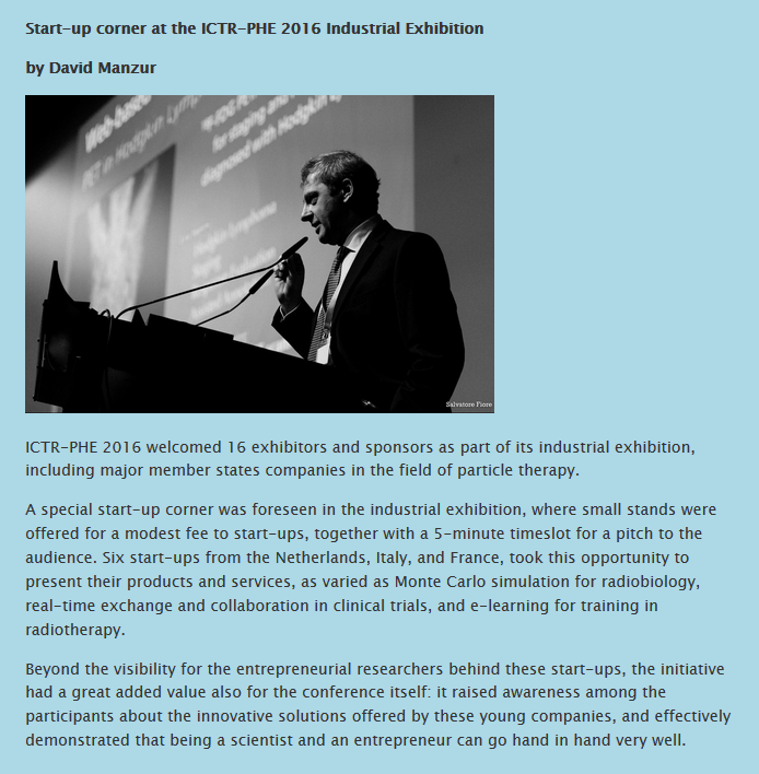
![]()
ICTR-PHE 2016: Uniting physics, biology and medicine
The third edition of the ICTR-PHE conference (see here), held in Geneva from 15 to 19 February, was again a unique place where multidisciplinary scientists met to share knowledge and to bridge gaps between all disciplines. More than 440 participants from all over the world met during five days and then returned to their home institutes with new ideas, new collaboration prospects, and optimistic visions of the future of cancer therapy.
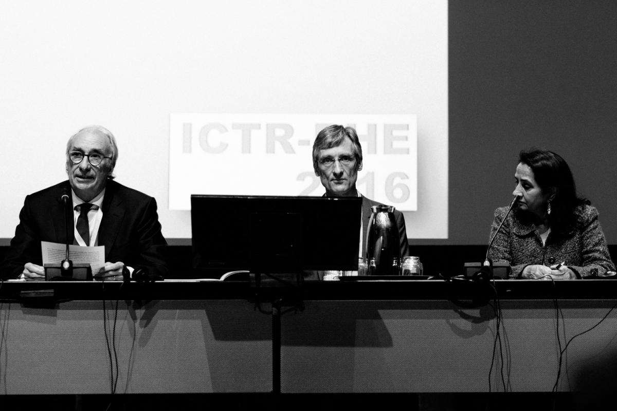
In their opening addresses on Monday 15 February, Eckhard Elsen, CERN’s Director for Research and Computing, and the conference chairs, CERN’s Manjit Dosanjh and Jacques Bernier from Clinique de Genolier, welcomed the participants, discussed the aims and how the primary scope of the event to “go from lab to bed” can be implemented effectively.
During five days, a very large spectrum of topics was covered – radiobiology, nuclear medicine, detectors and imaging, new technologies… – and many new researches were presented. This biennial conference is an important platform for representatives of the communities of physics, biology, and medicine, which have the occasion to gather, brainstorm and work together.
Indeed, already on that first day, experts in detector technologies, particle accelerators and nuclear medicine, as well as radiochemists, biologists and IT professionals were exchanging ideas and sharing their knowledge.
Some topics raised a particular interest in the audience, for example the studies on prompt gamma imaging, presented by Thomas Bortfeld, from the Massachusetts General Hospital and Harvard Medical School, and Saad Aldawood, from the Ludwig Maximilians University of Munich and the King Saud University of Riyadh. In particle beam therapy, the finite range of the beam can be a double-edged sword, because the over- or under-shoot of the beam requires extra margins in order to spare the healthy tissue. But this can compromise the dose distribution and the effectiveness of the therapy. That is why big effort is being put in developing imaging techniques for beam range assessment, and prompt gamma imaging appears to be the most promising one. It is based on the detection of secondary gamma radiation emitted from nuclear reactions of protons with tissue. It would allow detecting in real time the position of the beam in the body of the patient (during treatment) with an accuracy of about 1 mm. With such a precision, the range margins could be reduced, resulting in significant improvements of treatment quality.
The development and the clinical use of prompt gamma camera resulting from the ENVISION and ENTERVSION (projects that were funded by the EC and coordinated by CERN) was presented in detail by Christian Richter from Oncoray, Dresden.
Philippe Lambin, from the University Medical Centre of Maastricht (The Netherlands), tackled another promising topic, one we are very familiar with at CERN: big data. In a recent study, he shows how distributed learning can be a solution for rapid learning health care. “Rapid learning” is defined as the use of data routinely generated through patient care and clinical research to feed an ever-growing database. Thanks to this database, researchers hope to be able to develop mathematical models – following the example of weather models – capable of “predicting the future”.
The presentation on the use of magnetic resonance imaging (MRI) for external beam radiotherapy guidance, by Jan Lagendijk from the University Medical Centre of Utrecht (The Netherlands), also opened very new perspectives. Lagendijk explained that, although for certain tumours it is possible to have a good visualization of the cancerous structures with cone beam computed tomography linac (CT-linac) radiotherapy systems, it is not the case for all the tumours. On-line and real-time MRI guidance may offer the possibility to better imaging and thus targeting these tumors. This could provide a breakthrough in the application of radiotherapy and redefine the relation between radiotherapy and surgery.
In addition to senior and prestigious speakers, ICTR-PHE also featured the work of younger researchers: more than 100 of them presented their latest research in the poster sessions.

Finally, in line with its goal of merging different approaches and disciplines, the public talk by Domenico Vicinanza and Genevieve Williams, from Anglia Ruskin University in Cambridge, titled “Sound for Health – from Astronomy to Biomedical Sciences: Music and Sound as Tools for Scientific Investigation”, introduced a novel methodology that uses sound to improve our knowledge and understanding of human motor control.
To go further and have an overview of the 2016 ICTR-PHE presentations, you are invited to read the ICTR-PHE blog.